
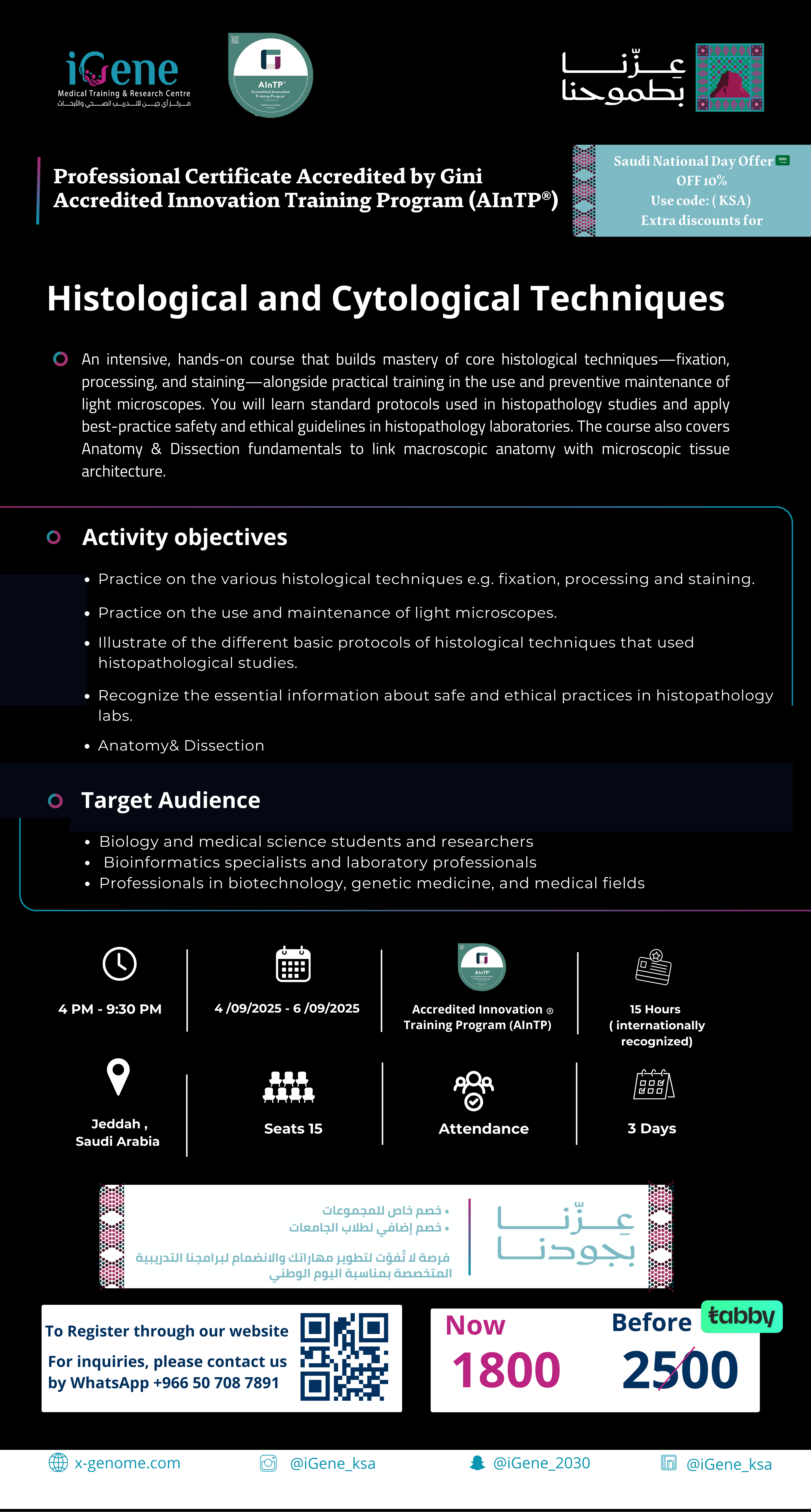
Histological and Cytological Techniques
“Registration has closed ”
Histological and Cytological Techniques
Price
1800 S.R
1800 S.R
Who is this course for?
Researchers, near-graduation and graduate students in the areas of Biological and Medical Sciences
Duration
15 hours (internationally recognized)
3 Days
certificate
Accredited Innovation Training Program (AInTP) ®
Location
iGene Medical Training & Research Center
Date
This course runs from Sep 11th, 2025, to Sep 13th, 2025
An intensive, hands-on course that builds mastery of core histological techniques—fixation, processing, and staining—alongside practical training in the use and preventive maintenance of light microscopes. You will learn standard protocols used in histopathology studies and apply best-practice safety and ethical guidelines in histopathology laboratories. The course also covers Anatomy & Dissection fundamentals to link macroscopic anatomy with microscopic tissue architecture.
Students will be able to:
1. Demonstrate proficiency and expertise in the proper use of the light microscope in examining histological specimens on glass slides after completing the course.
- Be familiar with the fundamentals of tissue fixation, dehydration, embedding, sectioning, staining, and mounting of slides for histological examination, immunofluorescent staining, and electron microscopy.
- Recognize, identify, and describe the characteristic structures of cells, tissues, and organ systems of the body using a light microscope, as well as digital microscopy and ultrastructural analysis for selected tissues.
- Know and understand the characteristics of body tissues (epithelium, connective, muscle, and nerve) and their relationships in the human body's various organ systems.
- Recognize the fundamental functions of cells, cellular organelles, tissues, and organ systems in relation to their histological structures.
- Recognize and comprehend the histological characteristics of selected tissues/organ systems as a result of disease processes (e.g., atherosclerosis, osteoporosis, pulmonary pneumonia, etc).
-
Anatomy& Dissection
Basic University training in Biological and Medical Sciences